Eye:
Dog Ear QuickLinks
Causes of Canine Glaucoma:
Glaucoma in dogs is a general term for a group of diseases, which causes increased pressure within an eye, resulting in an exhibition of severe symptoms due to the destruction of optic and retinal disks. It is estimated that around 1.7% of the dog population suffers from glaucoma. The condition is more common in those dogs with bulged eyes.
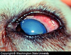
Picture Canine Glaucoma
Aqueous Humor; There is fluid in the normal canine eye which helps to maintain its shape, size and appearance. In order to maintain the correct level of intra-ocular pressure, the eye is constantly producing or eliminating fluid. “Aqueous Humor” or transparent fluids usually flows past the lens, through the pupil, across the inner cornea into the front part of the eye surface and is finally drained after maintaining the shape and consistency of the eyeball through fluid out flow. Since the cornea and lens do not have a blood supply, but they do need oxygen and nutritional supplies. This fluid, “Aqueous Humor” is also responsible for suppling the required level of oxygen and nutrition.
In some conditions, where the intra-ocular pressure becomes higher than normal for any reason such as an accident, trauma, inflammation, blockage, venous congestion etc, it causes damage to parts of the eye and as a result severe clinical symptoms are exhibited. Most commonly, optic and retinal discs are damaged and lens detachment causes partial to complete loss of dog eye function.
Types of Canine Glaucoma:
Dog glaucoma may occur due to any condition which causes in elevation of aqueous humor (Transparent fluid) levels in the eye ball. According to etiology or causative factors, canine glaucoma has two types, i.e. primary and secondary glaucomas.
Primary canine glaucoma; This type of canine glaucoma is noticed in certain breeds, which are genetically predisposed to the condition. Certain breeds such as Beagles, Hounds, Spaniels and Ekhounds etc, have a comparatively deficient eye anatomy, such as narrow drainage pores, a protruded eye globe etc.. These traits causes development of anatomical deformities/deficiencies in the eye globe which causes a primary type of increased pressure within an eye.
Primary canine glaucoma usually does not appear at once, but it develops with age, usually when a dog is 2 – 3 years of age. Similarly, primary canine glaucoma does not affect both eyes at once, but most commonly it consecutively causes pressure in dogs eyes, first in one and then the other.
Secondary canine glaucoma; This type of glaucoma in dogs is caused by some physical, pathological or unusual factors, resulting into a blockage and hindrance in the flow of aqueous humor (dog eye fluid). Different conditions and factors such as penetrating dog eye wounds (damage to the way fluids drain), inflammation (causes swelling), dog eye bleeding, lens displacement, scars over the iris, closed or narrow drain angles etc are some common dog eye problems that result in pressure that develops on the eye ball or glaucoma. Therefore, glaucoma may occur in cases where there are injuries, trauma, infections, chronic infections, inflammation and other pathological conditions which exerts pressure on the eye globe such as tumors etc.
Symptoms of Canine Glaucoma:
Dog eyes that develop glaucoma usually exhibit two forms of symptoms; acute and chronic. These two forms are of clinical importance and a treatment plan is usually selected on the basis of type and form of glaucoma found.
A dog may initially show signs of mild pain, minor to major irritation and swelling/protrusion of either of the eyes, where the white part of the eye seems to be bulged and veins across the sclera becomes prominent. The increased blood supply results from pressure and the white part of the eye may appear similar to “blood-shot-eyes” in humans. What is described here are common manifestations or symptoms. Dogs may not show all of these symptoms, but owners may surely suspect the developing condition as being canine glaucoma with either of these symptoms.
In advanced stages of this dog eye problem, the condition may worsen, causing severe pain, bulging and prominent signs in both of eyes. Major eye function, i.e. vision is lost partially or completely and other systemic problems appear such as a decline in the overall physiology of the dog (health).
Diagnosis of Canine Glaucoma:
Clinical signs which are described in taking a dog's history and detailed laboratory procedures are tools used to confirm glaucoma as a diagnosis and the exact cause of the condition.
Opthalmologic procedures and a detailed eye examination is required by a veterinarian, who will confirm the acuteness or chronicity (one or both eyes) of condition. The severity of glaucoma is also identified through clinical procedures.
Laboratory procedures such as tonometry, gonioscopy, ophthalmoscopy, electroretinograms and estimation of retinal and optic disk damage further confirms the severity and the exact cause of the canine glaucoma. Some or all of these procedures are usually required for confirmation, depending upon the nature, severity and chronicity of condition.
Treatment of Canine Glaucoma:
Once the type, form and cause of dog glaucoma is confirmed, treatment is done medically, surgically or with a combination of both approaches. In most cases, medical approaches to enhance the flow of aqueous humor is a short term treatment and cannot specifically treat underlying causes which are specifically primary in nature or where a condition is caused by an anatomical defect.
Infections which are causing inflammation can however be treated with specific antibiotics, anti-inflammatory drugs and steroidal eye drops and ointments.
Surgical correction of glaucoma in dogs is the ideal treatment, where anatomical defects, narrowness and blockage of drain tubes is done. Similarly, if possible, damage to optic and retinal discs are corrected with the help of complicated surgical procedures.
The treatment methods used have recently changed based on recent research which shows that a commonly used conventional treatment plan for canine glaucoma (combined surgical and medical approach), i.e. intrascelar prosthesis, enucleation (dependent surgical removal of the eyeball), cyclocryothermy (freezing procedure to reduce fluid production) and administration of antibiotics (gentamycin) and dexamethasone is no longer the treatment of choice because filtering tubules used tend to scar over again. For this reason, this has been proven to be short term treatment only.
For long term healing, a procedure forming anterior chamber shunts in the eye globe have proven to be very effective. Post surgical antibiotic and steroidal therapy along with the use of antifibrotic drugs prevents scaring and recurring blockage.
Symptoms such as pain, swelling and distress can be controlled by administering pain killers, anti-inflammatory drugs and the avoidance of bright lights. Similarly, bulged eyes and a protruded eye surface should be regularly cleaned with isotonic solutions. Natural remedies such as Eye Heal can also help to improve overall vision and health. Commercially available natural remedies can not only be used as post surgical supportive care but also regular usage can result in improved immunity, health and in strengthening of eye physiology. Others such as I-Clenz can be used to elan the face surface around the eyes to prevent infections.
|
|