Eye: Cataract
Dog Ear QuickLinks
Causes:
Canine eye cataracts are most commonly caused by a genetic or hereditary problem in some breeds. It is been noted that most dogs usually develop this problem at an early age. White terriers, English sheepdogs, Schnauzers, German Shepherds and Boston terriers can experience cataracts during the first few weeks of life. Similarly, American spaniels, Golden retrievers, Labrador retrievers, Rottweilers, Siberian huskies, Bull terriers and Standard poodles may develop this condition at age 6 – 12 months of age. Some breeds, like Bichon frise and Bay retrievers may develop this condition after 1 year of their age.
Other causes of dog eye cataracts may be aging, injuries, trauma, and radiation. Diseases that can cause cataracts as a secondary dog eye disease includes diabetes mellitus and various kinds of nutritional deficiencies.
A brochure from the American College of Veterinary Opthalmologists can be downloaded here.
Symptoms:
Congenital canine cataracts may occur at a very early age. As a result, the dog experiences vision difficulty. As the lens grows with age, the cataracts may be reduced as compared to the aperture of the lens, therefore restoration of eye function is noted. When the cataracts is a primary condition, the canine eye cataracts becomes less noticeable with age.
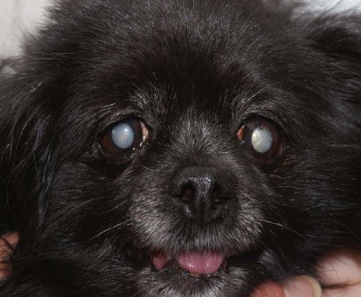
Picture Dog Eye Cataracts
Dogs suspected for eye cataracts should be examined for any change in eye coloration, vision problems and a reluctance to jump and walk in relatively darker environments. In some cases, symptoms such as inflammation, pain and partial to complete dog blindness may also be present. Such dogs should be suspected for cataracts and a veterinarian should examine it thoroughly.
Canine cataracts can appear as small spots, as a cracked-ice appearance, as a milky haze, as a white pearl sheen or white streaks. The dog eye cataracts can start is a small area and then expand across the dog eye lens. The rate at which the cataracts expands is difficult to predict.
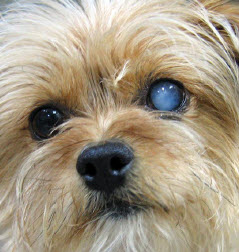
Picture Cataract in Dog
Eye
Cataracts can appear on 1 or 2 dog eyes
At the clinic, a veterinarian will examine the eyes after dilating the pupils. Specifically, cataracts appear as cloudy or opaque, like crushed glass or ice distributed on the surface of the eye. It effectively blocks light from reaching the retina.
Cataracts in dogs should be differentiated from nuclear density/nuclear sclerosis in older animals, where the lens appears to be smoothly darkened and gray in color, but not clouding. Similarly, dog cataracts should be differentiated from minor lens imperfections in young dogs, which is characterized by displacement and an anatomical disturbance of the eye apparatus.
Diagnosis:
Clinical examination and detailed breed information can help when identifying dog eye cataracts, but laboratory examinations such as detailed ophthalmology, retro-illumination, slit lamp bio-microscopy and optimal direct examination of the lens can confirm the anatomical distribution, location, degree of opacification and shape of the cataracts.
These procedures are also required for determining if the patient is suitable for a surgical treatment plan. Additionally, a detailed examination and laboratory procedures can also help when differentiating other conditions from cataracts such as the nuclear density of the lens and any lens imperfections caused by the cataracts.
Treatment:
When cataracts are removed surgically, a dog has to be clinically monitored and an owner has to carefully follow post-surgical instructions. Some dogs that undergo surgery may develop secondary canine uveitis (inflammation behind the cornea), which should be monitored. Supportive measures such as care and the use of natural remedies such as Eye Heal can be helpful, but are not a cure.
Video of Dog Cataracts Surgery
Dog eye drops are needed multiple times a day before a dog undergoes surgery and for 6 wks after the surgery is completed. Dogs that undergo treatment also need to wear an e-collar for 2 weeks. A dog must be brought in for checkups the day after surgery, and then at the 1, 3 and 6 week mark. According to the American College of Veterinary Ophthalmologists, surgery is successful 90% of the time.
In other dogs, in which the condition cannot be treated with surgery or where the condition is minor in nature, dog eye drops that are used that are formulated for dogs with cataracts. These dog eye drops are used for pupil dilation, antibiotics and other supportive components.
Dogs with cataracts need special care and training, through which they can live a quality life. Critical eye care, regular eye cleaning and the use of supportive eye drops such as natural remedies as mentioned above, can help to maintain overall dog eye health. Supportive eye drops and nutritional supplements can be used effectively along with the prescribed eye drops.
|
|