Digestive System: Liver Shunt
Types of Canine Liver Shunt Problems:
Congenital Canine Liver Shunt; Purebred dogs are primarily suspect and predisposed generically for the congenital type of live shunt. Breeds that are affected include Miniature Schnauzers, Terriers, Retrievers, Wolf hounds, German shepherds and Poodles. A congenital dog liver shunt either occurs extrahepatic (outside of the liver), or in different vessels such as the portal vein, splenic vein, gastric vein (Left), attachments of vena cava and in other systemic parts of the body that have to do with the blood supply.
Picture of Dog Liver
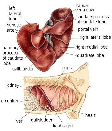
Picture of Dog Liver
The pictures in
this section are reprinted with permission by the copyright
owner, Hill's Pet Nutrition,
from the Atlas of Veterinary Clinical Anatomy. These illustrations
should not be downloaded, printed or copied except for personal,
non-commercial use.
Intraheptic canine liver shunt: This type of dog liver shunt is noticed within the hepatic blood supply. It occurs in younger dogs which are born with the fecal ductus closed at birth.
Acquired Canine Liver Shunt; This shunt is a chronic condition and common in aged dogs. It develops as a result of a hypertension in the portal vein. This type of liver shunt is more commonly noted in dogs that have severe and chronic hepatic (liver) disease; especially those diseases involving a large population of hepatic tissues, hepatitis, cirrhosis and fibrosis. It is considered that hypertension in the blood supply, especially in the portal vein and some other associated veins which supply blood to various systems occurs to prevent a fatal hypertension in the hepatic blood supply, which can lead to irreversible collapse and damage to the liver. Hypertension itself can cause the gradual developments of atrophy in the liver.
Symptoms of a Shunt Problem:
In either case, congenital or an acquired dog liver shunt, there is a strong possibility that hepatic encephalopathy (disturbances of consciousness that may progress to deep hepatic coma) can occur as a primary symptom. Other symptoms can include vomiting, diarrhea, lethargy, lack of muscular coordination, problems in growth, and weight loss or gain.
Problems with digestion are frequent due to a failure in the liver's ability to metabolize neurotoxins. A reduced ability to process blood makes things worse, i.e. the chances of having gastric ulcers, blood diseases and other mechanical circulatory disorders may occur.
Diagnosis of Canine Liver Shunt:
Symptoms are helpful in making an initial estimation of the condition, while laboratory procedures involving complete studies of blood content and a study of liver enzymes such as ALT, AP and BUN are required.
X-Rays on the other hand can help to estimate the status of the liver, the degree and location of the shunt. Abnormal anatomy and placement of the liver, ascites (fluid accumulation in the abdomen) and shunt location not only confirms the condition, but can also help to differentiate between the two different types of dog liver shunts.
Treatment of Liver Shunt:
An ideal option for treating a liver shunt in dogs is to perform surgical ligation of the shunt (closing or tyeing it off). This may be done as either a partial or complete ligation, depending upon the type and status of the abnormality. In most cases, if partial ligation is applied in the first surgery, an additional surgical procedure is usually required if clinical signs persist.
Some post surgical complications may occur, including bloody diarrhea, abdominal pain, shock, and cardiovascular collapse are usually noticed due to acute hypertension (high blood pressure) that occurs as a result of the ligation. Immediate medical attention for treating shock and removal of the ligation is indicated in such cases.
In cases where surgery is not possible, as in the case of post surgical complications or if the condition is diagnosed as minor, then medication is the treatment option of choice. Medication and management can help to reduce the severity of clinical signs, but it should be noted that it is never an alternative to specific surgical treatment, as liver atrophy progresses over time and the condition may worsen after some time.
Once the condition is treated completely, recovering dogs should be supplied with high energy diets combined with vitamins, supplements and herbal tonics, to hasten liver recovery and restoration. One homeopathic remedy that may be helpful is Liver-Aid Formula
Prognosis:
The prognosis for canine liver shunt is “favorable” if it is diagnosed and treated in time. The prognosis stands “less favorable” in dogs which have been treated with partial corrections or have experienced a delay in having the surgery done and kept on medications only.
|
|