Canine Cherry Eye
" Canine cherry eye is another name for dog eye prolapse and/or hypertrophy of the nictitans gland. It is more common in young dogs, but some breeds are predisposed. The exact cause of dog cherry eye is unknown, however it is considered to be a weakness in the connective tissues that are attached to the base of the dog eyes. Some tumors can also cause this condition. Cherry eye in dogs is characterized by swelling and dislocation of the third eyelid (nictitans gland and membrane), pus filled discharge, redness and signs of secondary infection. Treatment should not be initiated before confirmation of the cause and status of the dog eye condition. Treatment is either done by massage or surgical adjustment. Sometimes removal of the glands over the nictitating membrane is the only solution. In support, to reduce symptoms and routine eye care after treatment, natural remedies can help."
Causes of Canine Cherry Eye:
Unfortunately, the exact cause of this condition is unknown. It is more common in young dogs and breeds such as the American Cocker Spaniel, Beagle, English Bulldog and Lhasa Apso. It usually occurs in dogs under 2 years of age.
Some researchers believe that canine cherry eye is caused by weakness of the connective tissues which binds the nictitans gland and membrane to the base of the eye. Other researchers, have proven that dog eye tumors may cause the dislocation of the nictitans glands, thus it prolapses or hypertrophy occurs.
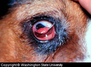
Picture of Cherry Eye in
Dog
Prolapse of the gland of the third eyelid (cherry eye) in a Beagle dog
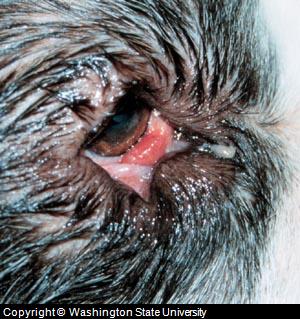
Picture of Canine Cherry
Eye
Prolapse of the gland of the third eyelid (cherry eye) in
a Saint Bernard dog
Symptoms of Canine Cherry Eye:
Symptoms of cherry eye in dogs are related to the stage of the condition. In the initial phases of this condition, the glands over the nictitating membrane get swelled and are dislocated from their original position. This is either by hypertrophy (dislocation due to swelling) or due to any pressure or weakness at the base of the third eyelid. In severe conditions, this gland appears as a red, compact body which sticks out of the membrane, over the surface of the front eye. That is why, this condition is called, “cherry eye”.
Due to a dog rubbing the eye and exposure of these glands to dust and pollutants, conditions can worsen and symptoms of extreme redness, puss filled discharge, swelling of the whole eye and secondary eye infections are noted. If the condition is left untreated, red swelling over the eye surface turns to a blackish color and starts to appear rotten.
Diagnosis of Canine Cherry Eye:
Although symptoms are always enough for identifying the dog cherry eye, it is essential that the exact cause and status of the condition be carefully assessed. Taking a dog's clinical history and careful examination of the eye is helpful in determining the status of the condition and the presence of any possible secondary infections, any tumors or any other unusual factor which caused the dislocation and swelling of nictitating glands from its membrane.
Lab tests are usually not required, but in severe and advanced cases of cherry eye in dogs, it might require culturing of any discharge and a biopsy in case a tumor is suspected.
Treatment of Canine Cherry Eye:
Since glands over the nictitating membrane are essential for the production of tears, which keeps the eye moist and clears the front eye surface, treatment of canine cherry eye should be done with care and after a thorough examination.
In the early stages of the disease and in mild cases, it can be done through massage. Both the upper and lower eyelids are closed with the fingers and with gentle massage, the membrane and glands are pushed towards the lower corner of the eye, towards the nose. Repeated massages can help in relocating the gland to its original cavity, but it is not always helpful and applicable, for the reason that either the case of dog cherry eye was reported late or the condition is not simple as it seems.
The most convenient way of treating cherry eye in dogs is surgical adjustment of the glands. Either the glands are repositioned surgically or in severe and irrecoverable cases, the glands are completely excised.
Repositioning of the glands might include suturing the glands with the nictitaning membrane or mucosal layers at the base of the eye. This is a successful treatment approach, however the physiology and anatomy of these glands are never termed normal, even after recovery. A course of antibiotics and supportive measures is essential after surgery.
If excision of the glands are required due to repeated occurrence of the canine cherry eye condition or if the condition is in an advanced stage, partial removal should be avoided as this might cause complications including severe secondary infections.
Dogs which are affected by cherry eye once in their life, even after recovery are more susceptible later in life to a dog dry eye condition and inflammation of the tear sac. Dog dry eye is certain in dogs at any stage of life, thus such dogs need continuous support for their eyes. This can be accomplished with regular eye washing and by using some naturally prepared eye solutions and drops such as Eye Heal to keep the eyes moist, clean, and in favorable health.
In addition, specific drugs and collagen supplements are used to improve the strength of connective tissues.
Have A Question or Helpful Story about a Dog Eye Problem ?
Have a Question, Request or Want to Share a Story that could help others? Our editors and pet health professionals will answer 1 question per week for free!
Please include information such as dogs history, description of the problem, dog age, breed, sex, Also include the dog's medical history in terms of past or recent diseases.
Including a picture of the dog eye problem is also helpful.
We will do our best to get back to you quickly (it depends on how many questions we receive each day). If you do require an immediate response we suggest using this online dog veterinary service that is available now.
Other Dog Eye Questions and Answers Submitted by Our Readers
Click below to see contributions from other visitors to this page...
What is this Dog Eyelid Bump? 
Maurice is a 9 /12 yr old great pyrenees with a bump on his upper eyelid. We have tried warm compresses 4 x daily. It has been about a yr. but does not …
|
|
References:
Merck Veterinary Manual (Merck & Co.)
Washington State University School of Veterinary Medicine


This site accepts advertising and other forms of compensation for products mentioned.
Such compensation does not influence the information or recommendations made.
We always give our honest opinions, findings, beliefs, or experiences.
All rights reserved. © 2018 Dog Health Handbook.