Dog Eye Anatomy
" Dog Eye Anatomy is a reflection of the eye as a complicated organ, containing many parts. These functional parts, i.e. sclera, conjunctiva, cornea, iris, pupil, lens, retina, lacrimal gland etc. are contained in a bony socket called a “orbit”. Along with these dog eye parts, muscles, blood vessels, nerves and tear drains are also part of the orbit, which play an important role in the normal functioning of a dog eye. Along with other dog eye problems, there are various conditions which are related to abnormal canine eye anatomy. These conditions are mostly treated with minor to major surgical procedures along with the use of specific therapeutics and supportive remedies, such as natural extracts to enhance recovery. ."
Dog Eye Anatomy Diagram
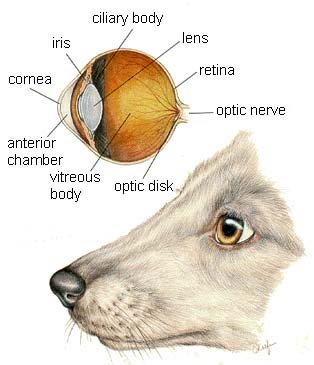
This picture is reprinted with permission by the copyright owner, Hill's Pet Nutrition, from the Atlas of Veterinary Clinical Anatomy. These illustrations should not be downloaded, printed or copied except for personal, non-commercial use.
Functional Layers of a Dog Eye:
On basis of function, the dog eye has three different layers:
- Fibrous Tunic: This part forms the outer most opaque layer and is comprised of collagen and fibrous tissues, called sclera. Sclera majorly forms the posterior of the eye, while anterior or the front part of the eye is the cornea, which is transparent and allows entry of light into the eye.
- Vascular Tunic: The dog eye is supplied with blood vessels, which supplies oxygen and nutrients to the all parts of the eye through the blood. The major part of the vascular tunic comprises a network of blood vessels, and also the choroid, which lies beneath the sclera, which helps in adjustment of the lens and “accommodation” or muscular constriction and relaxation which controls light. It is believed that this muscular mechanism is influenced by the vascular supply in the eye.
- Nervous Tunic: This functional part of the eye is controlled and is related to the nervous system of the dog eye anatomy. Photoreceptor cells called “retina” lies on the whole round wall of the posterior eye. These are controlled by retina, which collects and identifies the light, which is then processed to the brain with help of the “optic nerve”. The “Blind spot” or “Optic Disc” is the dark spot on the retinal wall, which has no photoreceptor cells over it and this part opens into the optic nerve duct, which contains a rich nerve supply, that connects the eye with the brain.
The aforementioned layers are functional layers of dog eye anatomy. The functionality of the eye is made possible with various anatomical features, which includes the, eyelids, eyelashes, conjunctiva, cornea, iris, pupil, lens, third eyelid (nictitans gland) lacrimal gland and viteous chamber of the eye.
The
following is a brief functional overview of these parts of dog eye
anatomy.
- Eyelids and Eyelashes: These are the outer most protective layers of the dog eye anatomy, which are flexible and wipe the tears over the eye surface to keep it protected and away from foreign particles such as dust and debris. The eye blinking mechanism is an involuntary action, which is controlled by the brain nerve supply. These parts also help to control light rays entering the eye.
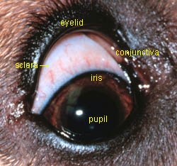
- Conjunctiva: Tough outer protective layers of the eye, the sclera is covered by a thin membrane, called conjunctiva. This thin membrane is located near the front of the eye. Conjunctiva runs over the cornea and covers all of the inner parts of the eyelids.
- Cornea: The cornea is the central dome shaped area of the eye surface, which is supposed to be a pathway for light entering into the eye. The cornea not only functions as a protective part of the inner eye but also it focuses light on the retina at the back of the eye. The focusing and light control function of the cornea is facilitated by the iris.
- Iris: This is the circular colored area of the eye, which controls the amount of light entering into the eye. Due to the functionality of this eye part, the pupil gets smaller or larger. The pupil is the central black area of the eye, which dilates or constricts under the influence of the iris and environment, to let more or less light entering into eye. The pupil dilates in a darker environment and constricts in a bright environment to control and allow an adequate amount of light to enter the eye.
- Lens: The lens lies just behind the iris of the eye, which is meant for focusing nearby or distant objects on the retina. The function of the lens is controlled by the ciliary muscles (small muscles) which contract, causing the lens to become thicker and nearby objects more focused. On the other hand, if the ciliary muscles relax, the lens becomes thinner and thus distant objects are focused on the retina.
- Retina: These are photoreceptor cells, which are distributed over the round back surface wall of the eye. The most dense part of the photoreceptor tissues or retina of the dog eye are called area centralis. In this region, thousands of photoreceptor cells are packed making the incoming images sharper. Each retina photoreceptor cell is attached with a nerve, which enters into the optic duct, through the optic disc and is connected to the brain. All the minor optic nerves of the retina are bundled into a larger optic nerve. Retinal tissues convert images into electrical impulses, which are carried to the brain with the help of the optic nerves.
- Nictitaing Membrane: In dogs, the eye is not only protected by the eyelids, but also with a nictitating membrane, which is also called the third eyelid. This is whitish to pink in color and is located on the edge of the inner part of the eyelids, near the nose. This membrane extends to the surface of the eye when the protection is needed or when the eye ball becomes vulnerable to scratches, irritation and in case of inflammation.
- Lacrimal Glands: These are tear producing glands. Tears keep the dog eyes wet and lubricated. Debris, dust etc is flushed out of the eye with the help of these tears. There are two types of Lacrimal glands, of which the lacrimal glands produce watery content or tears and the mucous glands in the conjunctiva produce mucous which mixes with tears to forms the protective nature of tears, as antibacterial or the slippery nature of tears wipe the debris out of the eye. Also the mucous part of the tears helps in preventing them from rapid evaporation. Tears are drained with the help of nasolacrimal ducts, which open into the nose.
Dog Anatomy and Dog Eye Health
In addition to a sound AAFCO certified diet (check the label),
a dog may benefit
from homeopathic remedies and dog eye cleaning during grooming to support dog eye anatomy.
Homeopathic remedies can be used to promote dog eye health,
reduce the
severity of symptoms during a dog eye problem and safely eliminate any
dog tear stain issues. Products used
should be from a reputable manufacturer and formulated specifically for
the Eye or surrounding areas. Two products that fit these
criteria include Eye
Heal which is the edges of the eye and
I-Clenz
which can help clean around the eye such as dog eye stains.
In terms of a dog eye wash, use an isotonic solution (isotonic means that the solution matches the biology of the dog eye) when needed such as the product offered by PNP. Many owners, as part of the grooming schedule will wash and check the eyes for any debris, infection or other issues. Other times a dog eye wash is used is to remove any chlorine, debris or salt water after swimming.
Have A Question or Helpful Story about a Dog Eye Problem ?
Have a Question, Request or Want to Share a Story that could help others? Our editors and pet health professionals will answer 1 question per week for free!
Please include information such as dogs history, description of the problem, dog age, breed, sex, Also include the dog's medical history in terms of past or recent diseases.
Including a picture of the dog eye problem is also helpful.
We will do our best to get back to you quickly (it depends on how many questions we receive each day). If you do require an immediate response we suggest using this online dog veterinary service that is available now.
Other Dog Eye Questions and Answers Submitted by Our Readers
Click below to see contributions from other visitors to this page...
Canine Glaucoma Can Progress to Blindness in 24 hours! Not rated yet
Our 8 yr old Saint Bernard, Clara is a goofy, spoiled member of our family. One Sunday morning, after attending church we noticed that she looked like …
|
|
References for Dog Eye Anatomy:
Merck Veterinary Manual (Merck & Co.)


This site accepts advertising and other forms of compensation for products mentioned.
Such compensation does not influence the information or recommendations made.
We always give our honest opinions, findings, beliefs, or experiences.
All rights reserved. © 2018 Dog Health Handbook.